The Muscular System and Hypertrophy
by Amy Bohan, MS | August 20, 2026
Case study video explaining the muscular system with a patient example on hypertrophic cardiomyopathy!
Use the button below to schedule a demo to learn about our quizzes, flash cards, and other anatomy resources that support this Brain Builder!
Schedule a Demo
by Amy Bohan, MS
The Muscular System
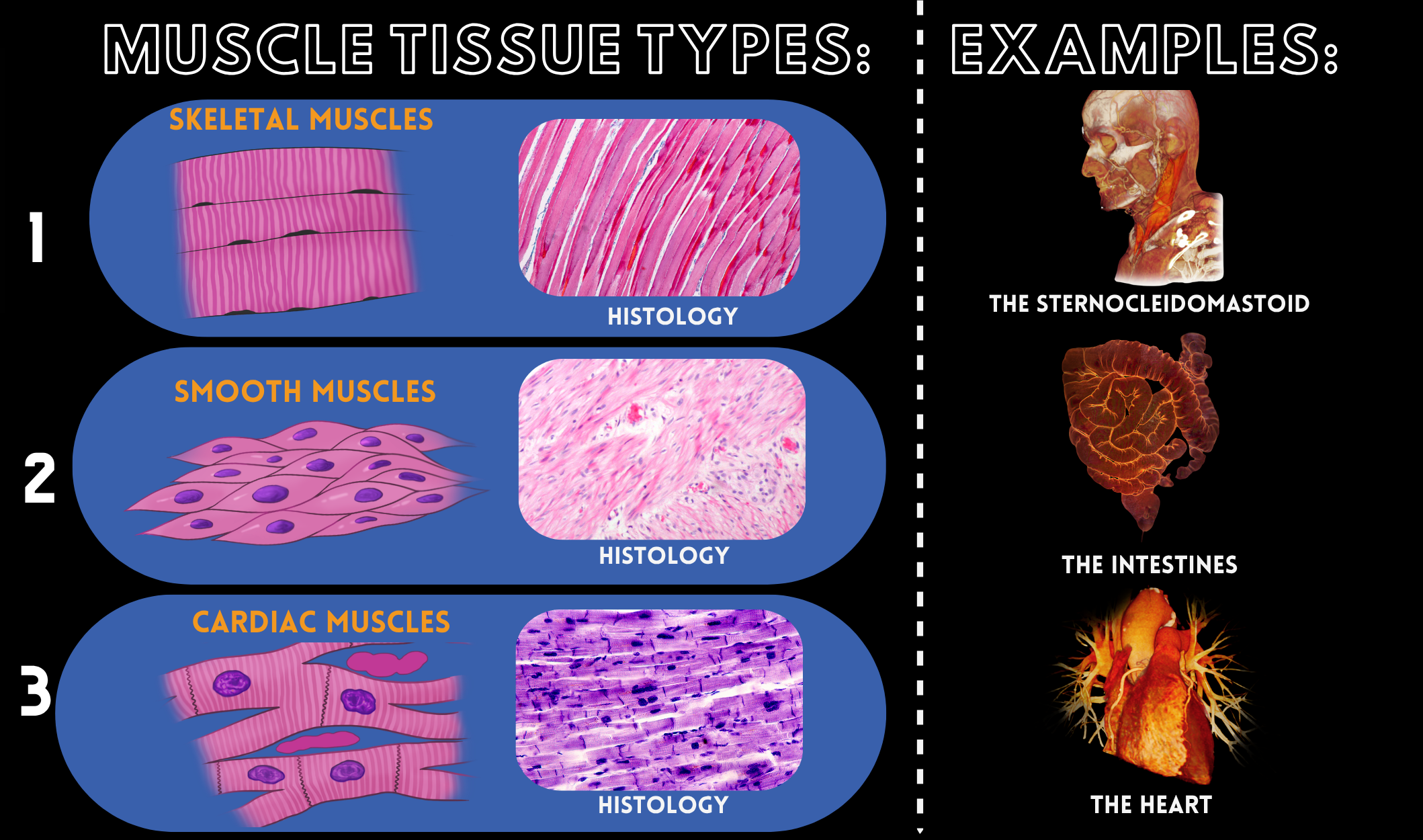
The muscular system consists of three subtypes of muscle: skeletal, smooth, and cardiac. Two of these (smooth and cardiac) function by involuntary movement, whereas skeletal muscle moves voluntarily. While there are some distinct differences, which we will explore below, there is one major similarity, and that is that all these muscles perform very specific functions and roles. Skeletal muscle is either directly or indirectly attached to bones, smooth muscle is found in many hollow organs and blood vessels, and cardiac muscle is found in the heart. I find it important to understand their structures on a microscopic level to best understand how these muscles undergo hypertrophy, hyperplasia, or both (in some cases). Let’s look at skeletal muscle and its components.
Skeletal Muscle
Skeletal muscle is composed of myofilaments, actin, and myosin, as well as troponin, tropomyosin, and titin. In addition, skeletal muscles and their nuclei are on the periphery of the parallel running fibers. All these filaments work together to help the skeletal muscle contract with the addition of calcium ions.
Cardiac & Smooth Muscle
Cardiac muscle has some components like skeletal, in that the striations are present in the fibers, which aid in contraction. However, we see the presence of intercalated discs in cardiac muscle, which aid in ensuring the cardiac pacemaker cells all contract simultaneously (think of a power strip). Furthermore, cardiac muscle has a branching appearance of the fibers, thus limiting its access to calcium, which is needed for contraction). The sarcoplasmic reticulum is located within these fibers, so shorter and branched fibers may limit this calcium availability. Cardiac muscle may, however, pull calcium from the extracellular fluid. Finally, smooth muscle has no striation, literally having a “smooth” appearance. Smooth muscle fibers appear tapered and have elongated nuclei.
Hypertrophy
Hypertrophy refers to growth in the actual size of the cell. We will examine what this means as far as these muscle types are concerned. Atrophy is the opposite of hypertrophy, and means “none” or “without growth”. One way to think of atrophy is to think of when one breaks a bone and must be in a cast, causing limited mobility for the individual. Inevitably, the affected muscle fibers will become smaller in size. Finally, the term hyperplasia refers to the increase in the number of cells (think mitosis or cell division) and proliferation. Since this article is going to focus mainly on hypertrophy, let’s examine the main causes of hypertrophy. The three main causes of hypertrophy are small tears in the muscle, metabolic stress, and muscle damage. *Betts et al.)
Hypertrophic Cardiomyopathy
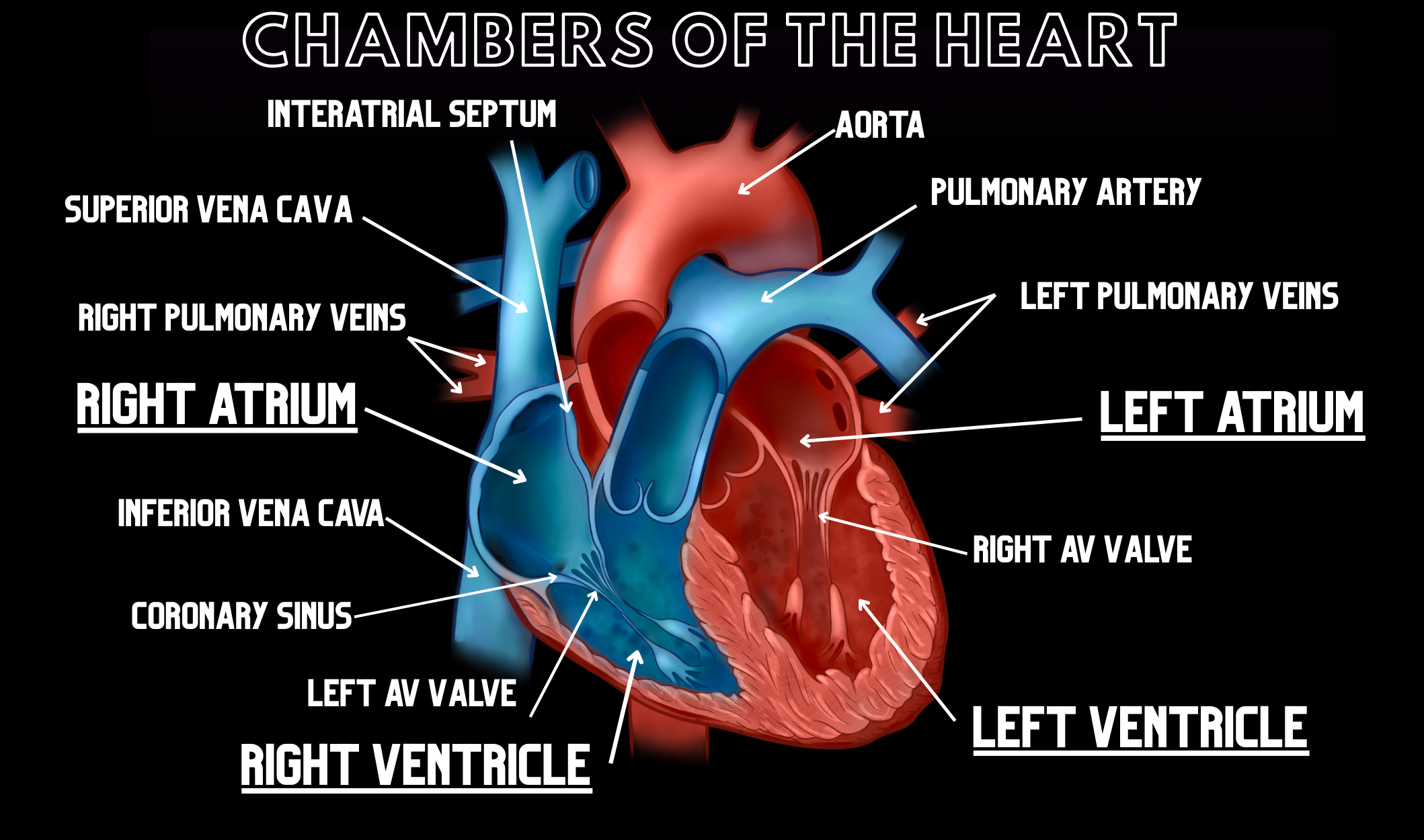
Now, let’s examine uncommon causes of hypertrophy, also known as homeostatic imbalances. There are two main examples , and the first is due to different neurological disorders. These diseases usually result in wasting, despite the name of hypertrophy. However, these are rare and sometimes, may not be as serious as the next example of homeostatic imbalance that we will discuss: hypertrophic cardiomyopathy (HCM). In HCM, there is a textbook thickening of the heart muscle (hypertrophy). Most times, cardiologists will diagnose this after witnessing a thickening of the ventricles as the heart muscle (myocardium), which are the lower two of the four chambers. In addition, the valves usually show some changes, as well, meaning that they may not be able to open and close correctly, thus allowing for the backflow of blood into the ventricles, compounding the problem. This can be particularly worrisome, because the left ventricle is the “pumping chamber” of the heart. If we think about the function of the left ventricle, it makes sense that symptoms could include shortness of breath, and dizziness. This is mainly due to the heart not being able to get blood out to the rest of the body. We need the oxygenated blood to help our tissues optimize at their maximum capacity. Therefore, one living with HCM may have a significant decline in their quality of life, compared to individuals unaffected by HCM. Oftentimes, diseases affecting the left ventricle can also lead to congestive heart failure. This could be life-threatening to individuals, and may be brought on by the heart muscle stiffening and hardening of the cardiac muscle.
Causes
The causes of HCM can vary, but genetics are often thought to be involved, as well as high blood pressure. Normal aging may even play a role. I would argue that the most dangerous part of HCM is that oftentimes, especially if left undiagnosed, it can lead to congestive heart failure. I find this most worrisome in the rare cases that someone isn’t experiencing symptoms of HCM at all, thus allowing this disease to progress into potentially more serious heart failure.
Diagnosis
A doctor may start diagnosing HCM by a multitude of tests, such as an echocardiogram, blood tests, and even x-rays (to check for heart enlargement). In some cases, an MRI may also be administered. (Betts et al.). Most likely, though, a medical professional would start the diagnosis process by examining the patient’s medical history and that of their family, since there appears to be some genetic component.
Treatment
Once a diagnosis of HCM has been given, many treatment options can prolong the quality of life. Beta-blockers and calcium channel blockers are first-line treatment options. As described at the beginning of this article, the presence of calcium ions is imperative to inducing muscle contraction. Therefore, the calcium channel blockers relax the heart muscle by not allowing so much calcium to be present. In addition, depending on the severity of the disease, one may even need an antibiotic to reduce the risk of certain bacterial infections of the endocardium (inner lining of the heart). (Betts, et al.). It is also imperative that one adjusts their lifestyle to their new diagnosis, meaning that they exercise with care. Changes in diet will most likely also need to be made, as one must limit water and salt intake. In rare cases, such as life-threatening ones, an implantable cardioverter defibrillator may be a procedure that is used. I think it is important to note that each patient is different, and a combination of some or all these treatments may need to be utilized.
Key Terms
Skeletal muscle - Voluntary muscle that is either directly or indirectly attached to bones; composed of filaments (actin and myosin) along with troponin, tropomyosin, and titin, with nuclei on the periphery of its parallel-running fibers.
Smooth muscle- Involuntary muscle found in many hollow organs and blood vessels. Smooth muscle has no striations, tapered fibers, and elongated nuclei.
Cardiac muscle - Involuntary, striated muscle found in the heart; its fibers have a branched appearance and contain intercalated discs.
Intercalated discs - Structures in cardiac muscle that help ensure the heart contracts as a coordinated unit by passing electrical signals quickly.
Sarcoplasmic reticulum - Found within muscle cells, this net-like organelle stores and releases calcium ions to control contraction and relaxation.
Hypertrophy - Growth in the actual size of a cell.
Atrophy - The opposite of hypertrophy, meaning “none” or “without growth.”
Hyperplasia - An increase in the number of cells (through mitosis/cell division) and proliferation.
Hypertrophic cardiomyopathy (HCM) - A pathological thickening of the heart muscle, most often diagnosed by thickening of the ventricles.
Schedule a demo today to learn how you can incorporate BodyViz into your classes and give your students the opportunity to practice using the information from this Brain Builder on real patients with authentic 3D dissection!
Schedule a Demo
Helpful Links: