Sleep Apnea
May 14, 2026

Case study video explaining the oral cavity, pharynx, and sleep apnea with a patient example!
Use the button below to schedule a demo to learn about our quizzes, flash cards, and other anatomy resources that support this Brain Builder!
Schedule a Demo
Overview
Although you may not have thought about it, the quality of your sleep is just as important to your cardiovascular and metabolic health as is a heart-healthy diet and regular exercise.
Research has concluded that uncontrolled sleep apnea, which is a condition in which breathing stops and starts several times during sleep, has been linked to heart disease and metabolic abnormalities, such as diabetes.
Often one’s bed partner may notice that their partner’s breathing pauses or stops during the night, or that their partner exhibits extremely loud snoring. Such observations may be an indication of sleep apnea.
However, it is important to understand that snoring is not the same as sleep apnea. Almost everyone snores at some time of their life. Snoring is the sound resulting from air passing over relaxed tissues within the nose and/or oral cavity.
Anatomy Related to Snoring and Sleep Apnea
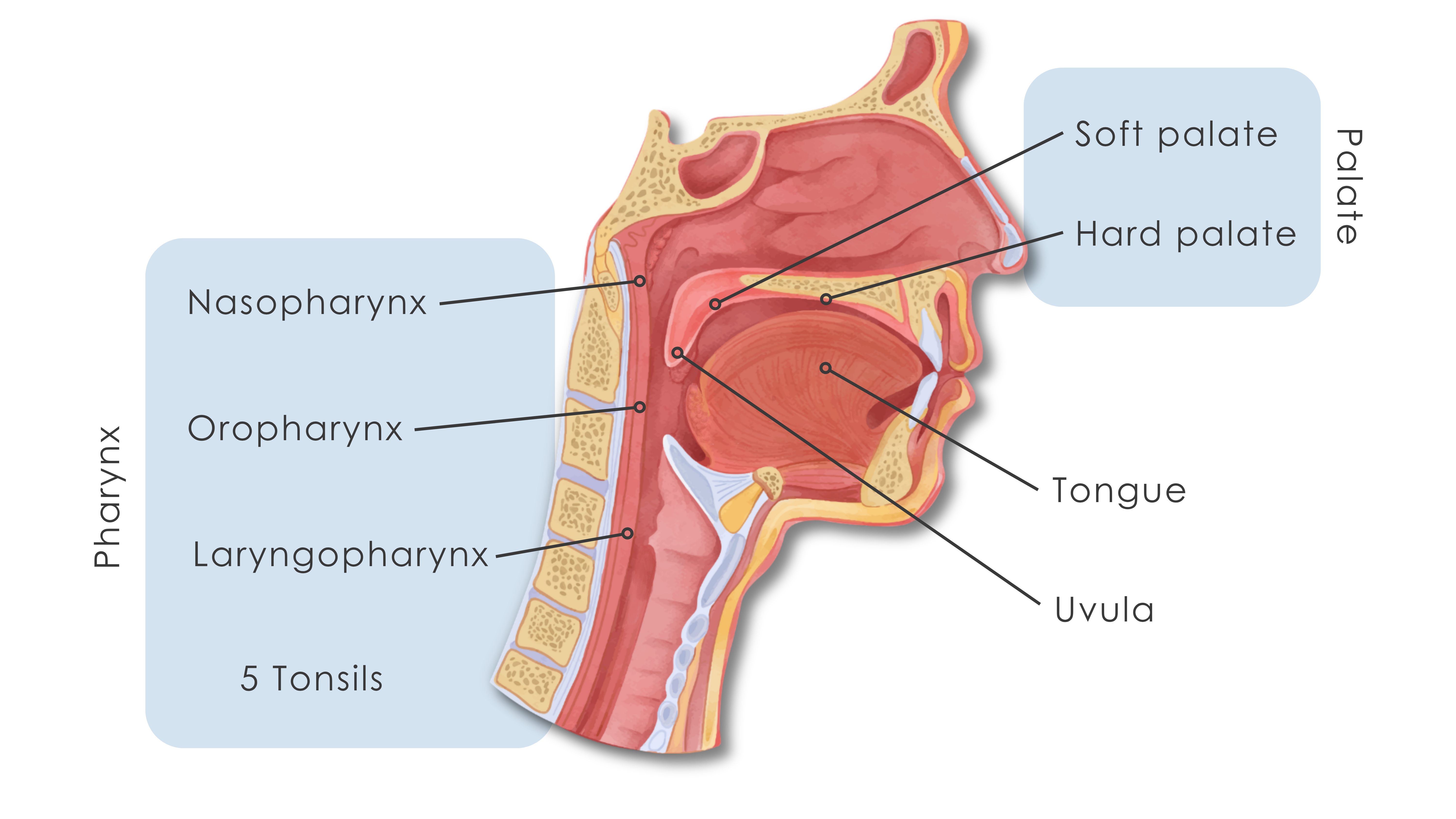
When one views a midsagittal section of the head and neck, the following structures, which are related to snoring and the more serious condition termed sleep apnea, are visible:
- The palate forms the arched roof of the mouth and the floor of the nasal cavity. It is composed of two segments: the hard palate and the more posterior soft palate.
- The soft palate is suspended from the posterior segment of the hard palate. The movable soft palate has no bony skeleton for support. Rather, it is composed of skeletal muscle and connective tissue. Laterally, the soft palate is continuous with the walls of the pharynx.
- The uvula is a conical, posterior extension of the soft palate that is visible hanging inferiorly within the posterior segment of the oral cavity.
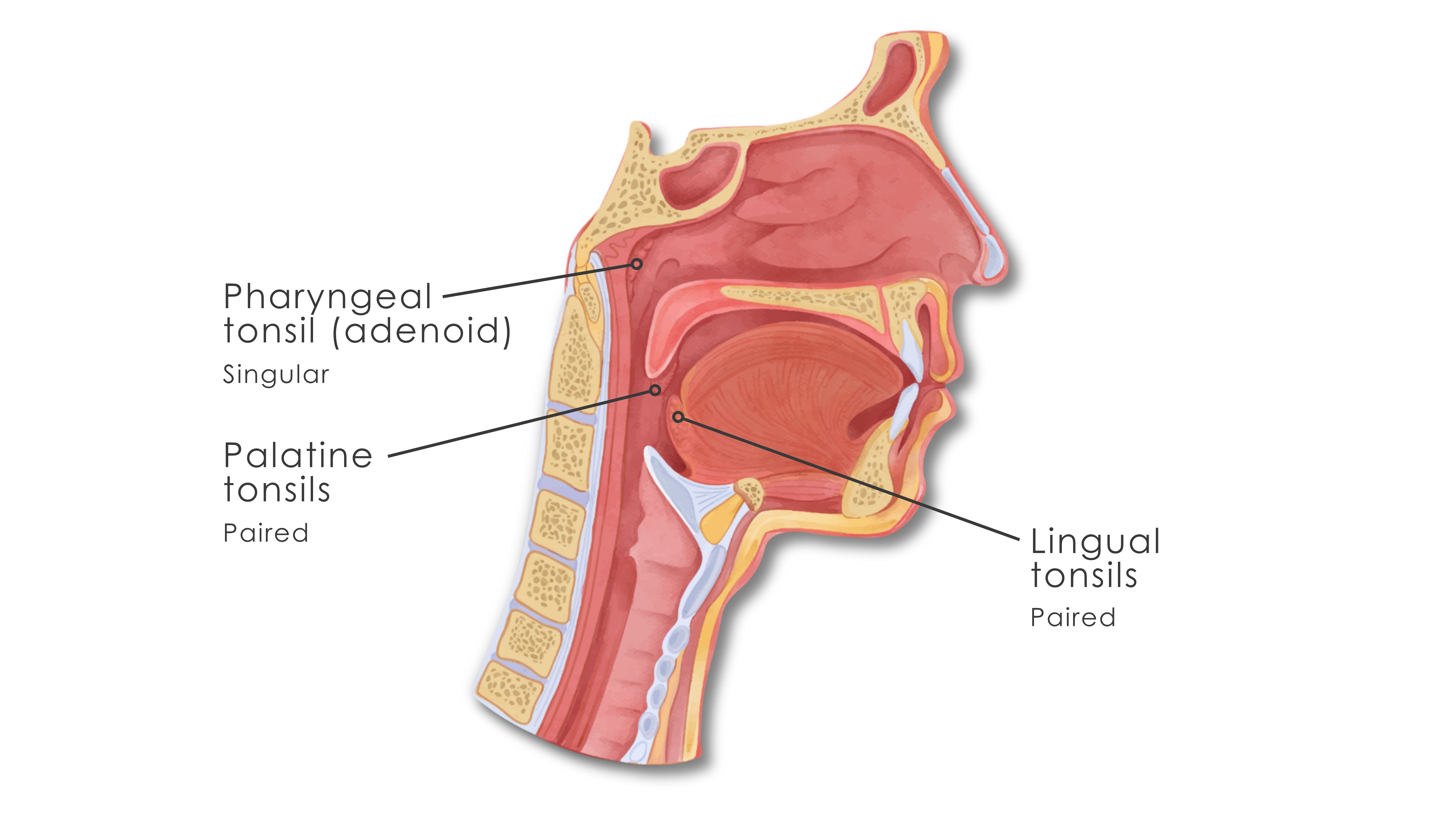
- The large lymphatic nodules in the walls of the pharynx are termed tonsils. The single pharyngeal tonsil, sometimes referred to as the adenoid, is located in the superior-posterior wall of the nasopharynx. The paired palatine tonsils are located in the inferior, posterior margin of the oral cavity along the boundary of the soft palate and the pharynx. The lingual tonsils are paired lymphatic nodules located deep to the mucosa at the base of the tongue.
- The tongue is an epithelial covered muscular organ that serves numerous functions related to the digestive system.
- The pharynx serves as a common cavity for the passage of air, solids and liquids.
Snoring results when air, passing over or past some of the tissues listed above, causes vibrations. These vibrations result in the sounds associated with snoring.
Lifestyle changes, such as a reduction in body weight, a change in sleep position (sleeping on one’s side), and a reduction in alcohol consumption close to bedtime may all reduce the frequency and volume of an individual’s snoring.
Signs of Sleep Apnea
If snoring is accompanied by one or more of the following signs or symptoms it would be advised to have the individual consult a physician in order to determine whether or not they have sleep apnea:
-
The observation by one’s sleep partner that breathing pauses during sleep
-
Restless sleep
-
Gasping or choking at night
-
Snoring so loud that it disrupts one’s bed partner’s sleep
-
Excessive daytime sleepiness
-
Difficulty concentrating
-
Increased incidence of headaches in the morning
-
Increased frequency of a sore throat upon awakening
-
High blood pressure
-
Chest pain at night
Sleep apnea has been found in approximately 3% of people exhibiting normal weight, and more than 20% of individuals that are obese. In addition, even though sleep apnea affects males more than females, the number of females exhibiting symptoms of sleep apnea increases significantly after menopause.
Types of Sleep Apnea
Clinically there are two types of sleep apnea
1. Obstructive Sleep Apnea (OSA) is characterized by repeated obstruction of the airway during sleep. Clinically, with this type of sleep apnea, the pharyngeal walls and soft palate narrow and collapse intermittently during sleep. The uvula and tongue may also move posteriorly. All of these movements obstruct the flow of air and the person stops breathing, resulting in interrupted sleep and snoring.
OSA can range from mild to severe. The degree of severity of OSA is based on a measurement termed the Apnea-Hypopnea Index (AHI), which measures the number of breathing pauses experienced within an hour. An individual with mild OSA will experience between 5 to 15 pauses per hour, while moderate OSA is characterized by 15 to 30 pauses per hour. An individual with severe OSA will experience more than 30 breathing pauses within an hour.
2. Central Sleep Apnea (CSA) is less common than obstructive sleep apnea, and often occurs in individuals who have another underlying health condition that affects the nervous system. Individuals with CSA experience poor-quality or restless sleep, and often wake up with shortness of breath or chest pain. Daytime symptoms, other than a lack of excessively loud snoring, are similar to those of OSA.
CSA is thought to be caused by a lack of appropriate brain signaling to breathe. This neurological abnormality is thought to be caused by the carbon dioxide sensors within the brain stem being less responsive to changes in blood levels of CO2.
Diagnosis
Diagnosis of sleep apnea is accomplished through a complete medical history, documentation of symptoms, a medical examination, and a diagnostic sleep study that determines the number of breathing events per hour.
Dangers of Sleep Apnea
There are a wide variety of complications that may result from sleep apnea including, but not limited to
- Cardiovascular problems: Blood oxygen levels drop repeatedly and suddenly occur during the breathing interruptions of sleep apnea. This can result in hypertension or significant stress on the cardiovascular system, sometimes resulting in a heart attack.
- Metabolic problems: Individuals with obstructive sleep apnea face a significantly higher chance of developing Type 2 diabetes as compared to individuals not experiencing sleep apnea.
- Complications during general anesthesia: Individuals with OSA are at a significantly higher risk for surgical complications during the use of general anesthesia.
Treatment
Treatment for sleep apnea is intended to resolve nighttime disruptions in breathing. This will improve the quality of sleep and reduce or eliminate possible health complications.
The primary treatment for sleep apnea is the prescription of positive airway therapy. Positive airway pressure therapy may be accomplished through the use of a variety of instruments during sleep.
- CPAP (Continuous Positive Airway Pressure) machine. This machine delivers air at a predetermined, consistent pressure to the individual during both inhalation and exhalation.
- BiPAP machine. This type of machine delivers air at one selected pressure at inhalation and a second, lower selected pressure, at exhalation.
- APAP (auto adjusting air pressure). This machine, which is the least frequently used machine, delivers air pressure at varying pressures in response to signals from the body, such as snoring or shifts in airflow.
Key Terms
CPAP - A continuous positive airway pressure machine that delivers air at a predetermined, consistent pressure during both inhalation and exhalation.
Obstructive Sleep Apnea (OSA) - This type of sleep apnea is characterized by repeated obstruction of the airway during sleep. Clinically, with this type of sleep apnea an individual’s airway narrows and collapses intermittently during sleep.
Central Sleep Apnea (CSA) - This type of sleep apnea is less common than obstructive sleep apnea, and often occurs in individuals who have another underlying health condition that affects the nervous system.
Snoring - The sound resulting from air passing over relaxed tissues within the nose and/or throat. Snoring may be caused by a variety of issues.
Uvula - A conical, posterior extension of the soft palate that is visible hanging inferiorly within the posterior segment of the oral cavity.
Adenoid - The single pharyngeal tonsil, sometimes referred to as the adenoid, is located in the superior-posterior wall of the nasopharynx.
Schedule a demo today to learn how you can incorporate BodyViz into your classes and give your students the opportunity to practice using the information from this Brain Builder on real patients with authentic 3D dissection!
Schedule a Demo
Helpful Links: