You Be the Doc: Developmental Anatomy and Real Patient Case Studies!
August 11, 2021
In contrast to a cadaver lab where the average age of a cadaver is 73 years-old, gross anatomy supplemented or supported by BodyViz gives students and instructors access to human anatomy at every stage of human development. In addition, BodyViz anatomy software allows students to see and explore anatomy from diverse backgrounds, medical histories, and life stages. In this anatomy case study, BodyViz gives you the opportunity to explore anatomy rarely found in a cadaver lab.
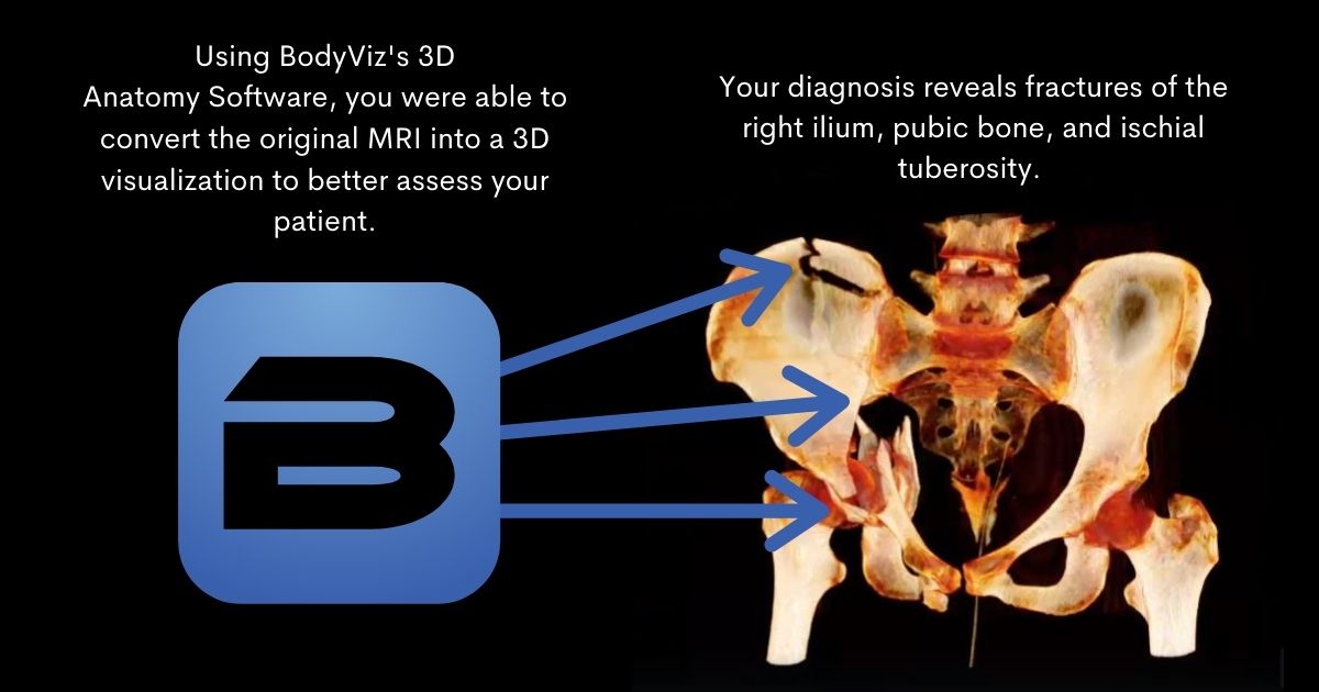
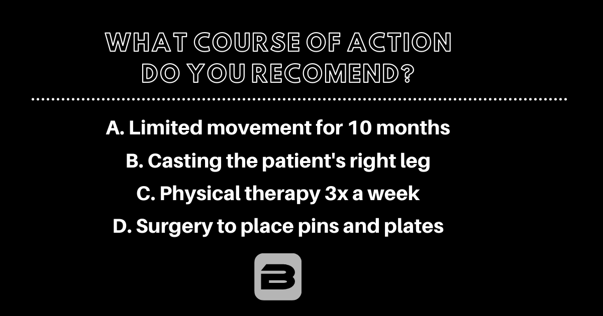
The real patient BodyViz Case Study features a 40 year-old female. Her symptoms include a swollen right hip with edema, bruising, and blood in her urine. During this “You Be the Doc” we asked viewers what course of action they would recommend after viewing the 3D dissection from the original CT scan of the patient. Take a look at the screen shots we have below, determine your answer, and then watch the video to see if you are correct!
Hint: What is the first step of action you would make?


Watch this video to see if you are correct!
Integrate BodyViz and Developmental Anatomy into Your Curriculum
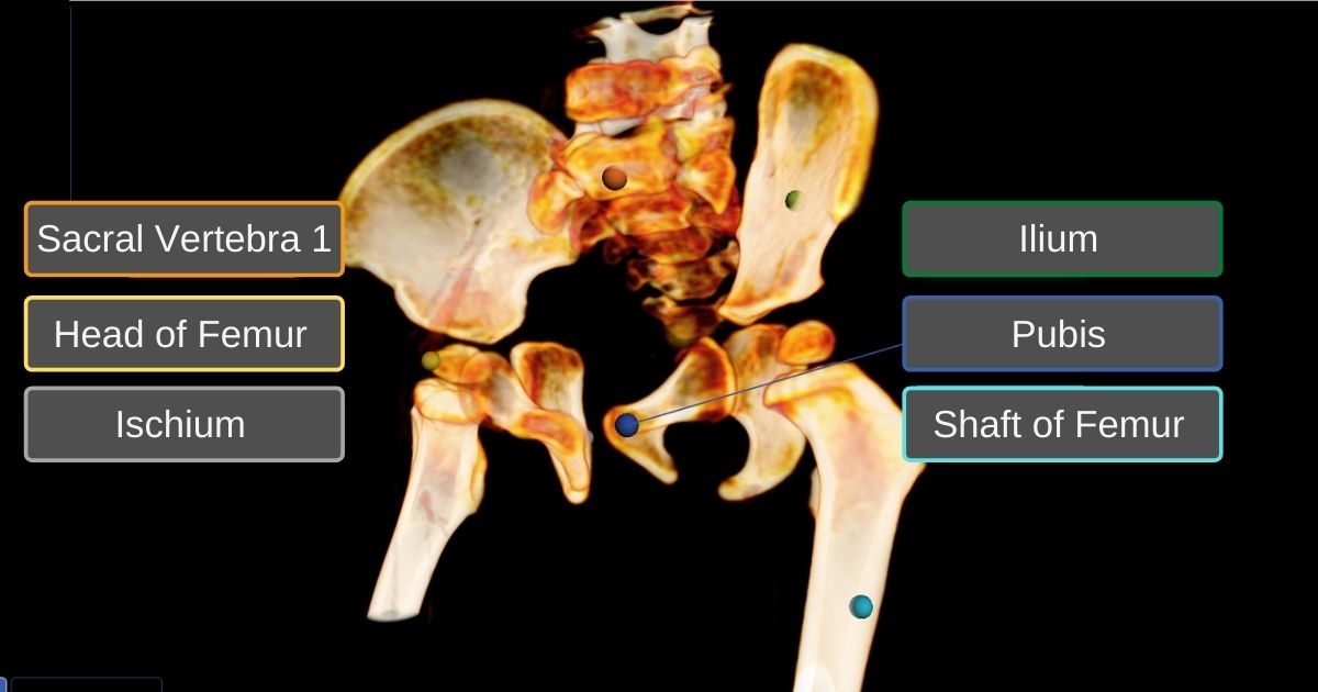
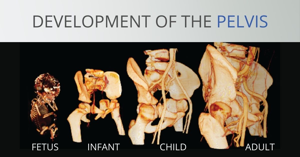
BodyViz also allows instructors to show stages of development. The following images are rendered from a CT scan of a 30-month old boy. The interactive 3D visualization focuses on the bones that make up the pelvis and hip joint. Over time the three bones—pubis, ilium, and ischium—will gradually fuse with the lower segments of the spine to form the adult pelvis and take on a solid, bowl-shaped form. In the leg, the head of the femur joins with the shaft of the femur to make the familiar ball-and-socket joint of the acetabulum in the hip. In this 3D CT visualization, we get a rare view of these separate bones in the developing toddler. Understanding how these bones originate and how they form together is critical when discussing many developmental pathologies.
The following images are screenshots from two different anatomy courses. Bringing real and diverse anatomy into your classroom will increase student engagement and class comprehension as students are able to apply anatomy to real life examples. BodyViz easily integrates into current curriculum and LMS systems, supplements gross anatomy lab, supports problem based learning, and provides on demand access to real anatomy.
Learn more about BodyViz solutions here.


With BodyViz, the learning possibilities are limitless! Because BodyViz 3D anatomy dissection software converts MRI and CT scans into 3D visualizations, users are able to upload their own MRI and CT scans on top of the 1000+ scans BodyViz already has!
Learn more about how BodyViz makes all stages of human development and real patient studies accessible to anatomy instructors and students using the links below and Scheduling a Demo!
Schedule Demo
Helpful Links