How Virtual Dissection Tables Support Preoperative Teaching
July 21, 2026
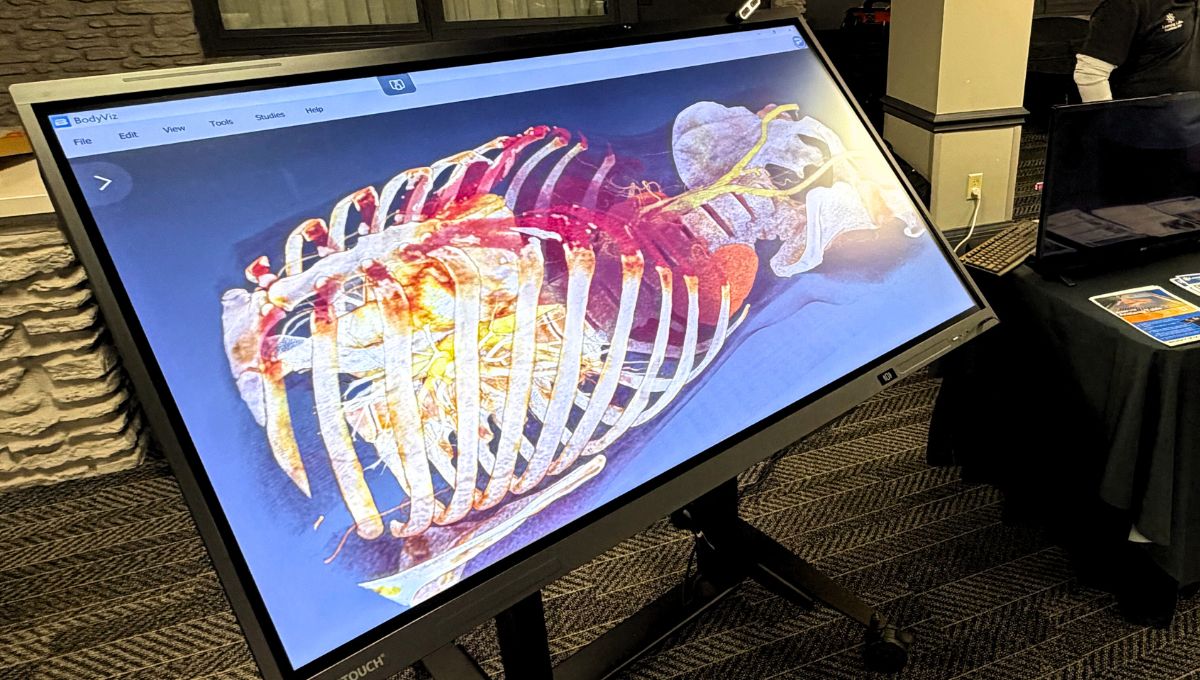
Preoperative anatomy teaching requires more than textbooks and static images. When medical students and residents need to understand how anatomical structures relate to surgical approaches, they need tools that reveal spatial relationships in ways that traditional methods often cannot. Virtual dissection tables have emerged as valuable instruments for connecting foundational anatomy knowledge with clinical case preparation, giving instructors new ways to demonstrate the structures their students will encounter in surgical settings.
This article explores how virtual dissection technology supports preoperative anatomy teaching, why clinical case correlation matters for student preparation, and what instructors should consider when integrating these tools into their curricula.
Key Takeaways: How Virtual Dissection Tables Support Preop Teaching
- Virtual dissection tables allow students to explore anatomical structures in 3D, building the spatial reasoning needed for surgical planning.
- Clinical case correlation becomes more accessible when instructors can display real patient imaging alongside normal anatomy.
- BodyViz enables instructors to upload MRI and CT scans, creating personalized learning experiences tied to specific surgical scenarios.
- Repeatable virtual dissections allow students to practice identifying structures without the time constraints of cadaver lab sessions.
- Access to over 1,000 real patient scans helps students recognize anatomical variations they may encounter during clinical rotations.
What Are Virtual Dissection Tables and How Do They Work?
Virtual dissection tables are interactive platforms that display 3D reconstructions of human anatomy derived from real MRI and CT scan data. Students interact with these visualizations through touch screens, adjusting views, removing layers of tissue, and examining structures from multiple angles.
Unlike static anatomical atlases, these tools let learners rotate specimens, zoom into specific regions, and reset to original states for repeated study. Instructors can guide students through virtual dissections while highlighting surgical landmarks and demonstrating how structures relate to one another in three-dimensional space.
Why Spatial Reasoning Matters for Preoperative Teaching
Surgeons operate in three dimensions, navigating complex tissue planes and anatomical relationships that textbooks cannot convey effectively. Students who develop strong spatial reasoning during their preclinical years are better prepared to interpret what they see during procedures.
A 2023 study published in Advances in Medical Education and Practice found that medical students using virtual anatomy modules showed statistically significant improvements in their understanding of anatomical relationships. The research demonstrated that 91.2% of participants improved their assessment scores after completing modules that incorporated 3D visualization with clinical case studies.
How Virtual Dissection Tables Support Clinical Case Correlation
Connecting anatomy knowledge to clinical presentations is essential for preoperative teaching. When students can visualize both normal anatomy and pathological conditions side by side, they begin to understand how diseases affect structures and how surgical interventions address those changes.
Instructors using BodyViz can display real patient scans that show conditions students will encounter in their clinical rotations. By examining examples of aortic aneurysms, vertebral fractures, or cardiac abnormalities in 3D, students develop pattern recognition skills that transfer directly to surgical planning contexts.
Building Connections Between Imaging and Anatomy
Modern medicine relies heavily on diagnostic imaging, yet many anatomy courses have historically separated imaging instruction from dissection. Virtual dissection platforms bridge this gap by presenting anatomical structures alongside the radiographs used to identify them clinically.
Students learning to read CT and MRI images benefit from seeing how cross-sectional views correspond to the three-dimensional structures they represent. This integration helps future physicians interpret imaging studies with greater confidence when preparing for surgical procedures.
What Are the Limitations of Traditional Cadaver Labs for Preoperative Teaching?
Cadaver dissection remains valuable for understanding tissue texture and developing manual skills, yet it presents challenges for preoperative anatomy instruction. Specimens are fixed in a single state, cannot demonstrate anatomical variation across multiple bodies, and deteriorate with use.
Access issues compound these challenges. A 2025 review in 3D Printing in Medicine highlighted that many institutions face cadaver shortages, limited lab hours, and rising costs associated with specimen acquisition and preservation. These constraints can reduce the time students spend with anatomical structures before entering clinical settings.
Virtual dissection tables address several of these limitations by offering repeatable access to diverse patient examples without degradation or scheduling restrictions.
How Do Instructors Integrate Virtual Dissection into Preoperative Curricula?
Effective integration begins with curriculum mapping that aligns virtual resources with specific learning objectives. BodyViz partners with instructors to create customized implementation plans that match their course schedules and teaching goals.
The curriculum mapping process starts when instructors share their syllabi, course schedules, or lesson plans. Content specialists then identify appropriate 3D scans, interactive modules, and assessment activities from an extensive library of real patient data. This approach reduces instructor prep time while ensuring students engage with relevant material at the right points in their training.
Supplementing Rather Than Replacing Traditional Methods
Virtual dissection works best as a complement to other teaching approaches rather than a complete replacement for hands-on experience. Students benefit from the tactile feedback of cadaver dissection while using virtual tools to preview structures, review material, and explore additional examples.
The American Medical Association has noted that virtual anatomy labs won't fully replace traditional dissection, but they can significantly enhance student preparation and reduce uncertainty when students enter cadaver labs for the first time.
What Features Should Anatomy Instructors Look For in Virtual Dissection Technology?
When evaluating virtual dissection platforms for preoperative teaching, instructors should consider several factors that affect educational utility and ease of implementation.
Platforms built from real patient scan data offer advantages over those using idealized computer-generated anatomies. No Two Bodies are Alike, and students preparing for clinical work need exposure to real natural variations that they will encounter in actual patients. BodyViz gives instructors access to over 1,000 real MRI and CT scans showing anatomical variation, pathology, and developmental stages across patient populations.
Instructors should also evaluate how well a platform supports clinical case correlation. The ability to display real patient imaging, upload institution-specific MRI and CT scans, and compare normal anatomy with pathology helps students connect foundational anatomy to the clinical scenarios they will encounter during rotations and future practice.
Ease of implementation is another important consideration. Platforms that include curriculum mapping, instructor training, and ongoing support reduce preparation time and make it easier to integrate virtual dissection into existing courses. Rather than building lessons from scratch, instructors can begin teaching with resources already aligned to their learning objectives.
Finally, flexibility is essential. Virtual dissection platforms should support learning across lectures, laboratories, and independent study while giving students access on multiple devices. This allows learners to review anatomy before class, reinforce concepts after lab sessions, and continue studying outside scheduled instructional time.
Flexibility Across Learning Environments
Effective platforms work across multiple settings. Students should be able to access materials during scheduled lab sessions, in lectures, and during independent study time. Mobile compatibility withthe BodyViz Portal extends learning beyond classroom walls, allowing students to review before exams or prepare for upcoming clinical experiences.
How Does 3D Visualization Improve Student Comprehension and Retention?
Research supports the educational value of three-dimensional visualization in anatomy courses. Students often find it difficult to construct mental models of spatial relationships from two-dimensional images alone, particularly when studying complex regions like the head, neck, and pelvis.
Virtual dissection tables allow students to manipulate structures, view them from surgeon-relevant perspectives, and immediately see how components fit together. This active engagement tends to produce better retention than passive observation of static images or even witnessing instructor-led cadaver dissections from across a crowded lab.
Preparing Students for Clinical Rotations and Surgical Observation
The goal of preoperative anatomy teaching extends beyond examination performance. Students who can quickly orient themselves to surgical anatomy, identify key landmarks, and understand procedural approaches contribute more meaningfully to clinical teams.
Virtual dissection platforms help build this readiness by exposing students to the same imaging modalities and anatomical perspectives used in surgical planning. BodyViz enables instructors to upload institution-specific cases, creating direct connections between classroom learning and the clinical cases students will encounter during rotations.
FAQs about How Virtual Dissection Tables Support Preop Teaching
Can virtual dissection tables completely replace cadaver labs?
No, virtual dissection tables supplement rather than replace cadaver-based instruction. Hands-on dissection develops tactile skills and familiarity with tissue properties that virtual tools cannot replicate. Combining both approaches gives students the most complete preparation for clinical work.
What types of clinical cases can be taught using virtual dissection?
Virtual dissection platforms support teaching across most anatomical regions and clinical scenarios. BodyViz offers modules covering cardiovascular conditions, abdominal pathology, musculoskeletal injuries, and neuroanatomy, all visualized from real patient scan data that instructors can customize for their curricula.
How do virtual dissection tables help students prepare for surgical procedures?
These platforms allow students to visualize surgical approaches from multiple angles, identify key landmarks, and understand how anatomical structures relate during procedures. BodyViz enables instructors to walk students through preoperative planning steps using the same imaging data surgeons review before operations.
Are virtual dissection platforms accessible outside of scheduled lab times?
Yes, platforms like BodyViz offer 24/7 access through student downloads and portal access. Students can review anatomy content on personal devices, including through the BodyViz iPad App, allowing study to continue anytime and anywhere that fits their schedule.
How does BodyViz support instructors implementing virtual dissection technology?
BodyViz partners with instructors every step of the way through curriculum mapping, training sessions, and ongoing support. Content specialists create customized implementation plans aligned with course objectives, reducing instructor prep time while ensuring students engage with appropriate 3D anatomy content.