Why No Two Bodies Are Exactly Alike
September 2, 2025

When we teach human anatomy, we often present a standardized map of the body with every artery, muscle, and bone exactly where it’s “supposed” to be. But the reality that healthcare professionals find as they head into the medical field for the first time, is that no two bodies are identical. Understanding anatomical variations is essential for students who aim to practice healthcare, to provide accurate diagnoses and effective treatments. From subtle differences in vascular pathways to significant structural anomalies, the human body is full of surprises that can challenge even the most experienced clinicians.
In this article, we’ll explore what anatomical variations are, common variations and why these differences matter, and how BodyViz is revolutionizing how students and educators explore anatomical diversity.
What Are Anatomical Variations?
Anatomical variations are natural differences in the structure of the human body. These differences aren’t necessarily pathological or abnormal, they’re simply alternative expressions of human anatomy that fall within the normal range of human diversity.
Professor Greg Smith from Saint Mary’s College of California, who has been teaching anatomy since 1981, explains:
“BodyViz allows the students to really see an object as though it’s in front of them, no matter how they manipulate it. This is crucial for understanding anatomical variations that textbooks simply can’t adequately represent.”
Some people are born with an extra artery near the kidney (accessory renal artery), while others might have variations in their biliary tract or venous drainage patterns. These changes may never cause problems, but in some cases, they can significantly influence how a condition is diagnosed or treated.
For future healthcare professionals, being aware of these variations can be the difference between a routine procedure and a serious complication. Students who learn with 3D anatomical resources demonstrate improved spatial understanding of complex structures and their variations.
Why These Differences Happen
Every body is shaped by a complex interplay of genetics, environmental factors, and evolutionary history. Virtual anatomy dissection tools allow students to explore human structures from any angle, revealing variations that might otherwise go unnoticed.
Several factors contribute to anatomical variations
-
Genetic Diversity: Slight changes in DNA can lead to structural differences in how the body develops.
-
Developmental Factors: During embryonic development, slight alterations in timing or signaling can result in anatomical variations.
-
Evolutionary Adaptations: Some variations represent evolutionary adaptations that may have provided advantages in certain environments.
-
Environmental Influences: Factors during development, including maternal nutrition and environmental exposures, can influence anatomical development.
The result of these factors is that no two people, not even identical twins, have exactly the same anatomical structures. This diversity is both a challenge and an opportunity for medical education.
The Clinical Significance of Anatomical Variations
Understanding these differences isn’t just academically interesting, it’s critical in clinical settings. Studies show that interactive anatomy learning leads to better knowledge retention and clinical application.
Dr. Bob Frampton, who uses BodyViz in his physical therapy program, emphasizes:
“Today’s students don’t want to be talked to. They want to be engaged in a conversation. BodyViz creates that interactive environment where students can explore anatomical variations and directly relate them to patient care scenarios.”
The clinical implications of anatomical variations are vast
-
Surgical Planning: Surgeons need to know where a blood vessel might be, not just where it should be. Variations in vascular anatomy can dramatically alter surgical approaches.
-
Diagnostic Accuracy: Radiologists must be able to distinguish between normal variations and pathological findings. What might look abnormal could simply be a common variation.
-
Procedural Success: Even routine procedures like central line placement or regional anesthesia can be complicated by anatomical variations.
-
Treatment Efficacy: Variations can affect how medications are distributed throughout the body or how certain treatments work.
When these variations are overlooked, it can lead to misdiagnosis, surgical errors, or delayed treatment. That’s why incorporating these topics into anatomy education helps students become more confident and better prepared for real-world clinical scenarios.
Common Variations You Might Not Learn from a Textbook
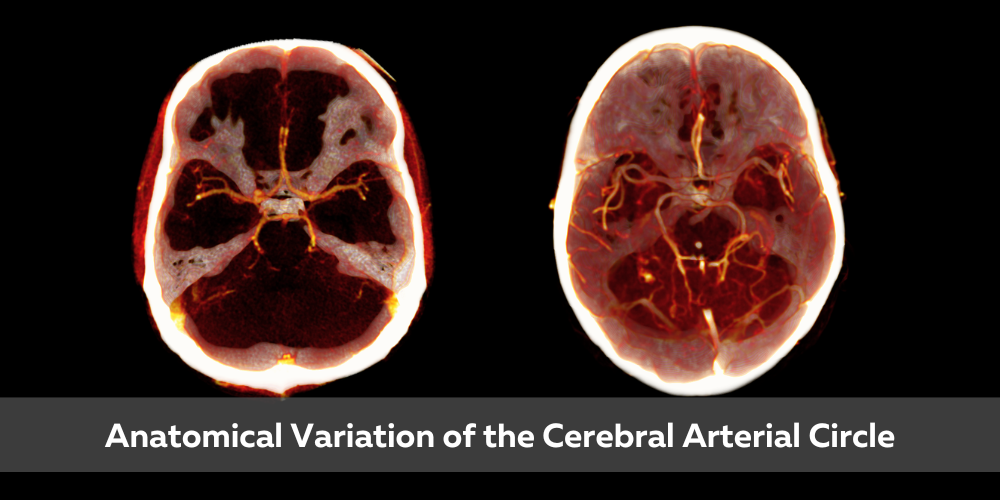
Some of the most significant variations occur in the vascular system. For example, the Circle of Willis, an important network of arteries at the base of the brain, can look completely different from one person to another. These differences can affect how a stroke is diagnosed or how the brain is supplied with blood.
The best anatomy software for educators combines ease of use with comprehensive visualization capabilities to demonstrate these variations effectively.
Notable Anatomical Variations
Vascular Variations
-
Up to 30% of people have variations in the branching pattern of the aortic arch
-
Accessory renal arteries occur in approximately 30% of individuals
-
Variations in hepatic arterial supply affect up to 45% of the population
Muscular Variations
-
About 15% of people don’t have the palmaris longus muscle in their forearm
-
The psoas minor muscle is absent in approximately 40% of individuals
-
Variations in facial musculature can affect facial expressions and surgical outcomes
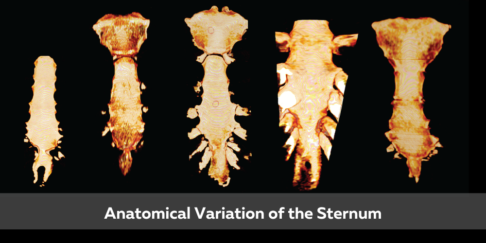
Skeletal Variations
-
About 1 in 200 people have an extra cervical rib
-
Variations in vertebral anatomy occur in up to 12% of the population
-
The number of lumbar vertebrae can vary from 4 to 6
Nervous System Variations
-
The course and branching of peripheral nerves can vary significantly
-
Up to 20% of people have variations in the brachial plexus
-
Cranial nerve pathways can follow alternative routes
These kinds of differences might not be noticed until a medical issue arises or until a student discovers them during anatomy training. Virtual dissection tools allow for repeated practice without the limitations of traditional cadaver labs.
The Limitations of Traditional Methods
Traditional anatomy education faces significant challenges when it comes to teaching anatomical variations. In a typical anatomy lab, students might work with one or two cadavers throughout their course. This limited exposure means they might never encounter important variations that could be clinically relevant in their future practice.
Amy Wuthrich, an experienced nurse and educator who chairs DeSoto County High School’s CTE program, initially faced this challenge:
“I’ve spent the last six years finally honing my toolkit for the class. And that’s why I’m so excited about BodyViz. It gives them something they can see, something tangible that they can play with and manipulate.”
Textbooks and atlases typically show the “classic” or most common anatomical arrangements. While some may mention variations in footnotes or supplementary sections, these static images don’t allow students to fully visualize or interact with these differences.
Even when variations are discussed, traditional 2D images make it difficult for students to understand the spatial relationships between structures, which is crucial for clinical application.
Dr. Terry Maksmowych, an experienced educator, was initially skeptical about virtual anatomy tools:
“My first thought was ‘Oh, I’ve heard of those types of things at conferences and they’re so expensive that there’s no way we could afford doing that.’ But BodyViz has proven to be both accessible and incredibly effective for helping students visualize anatomical relationships.”
Not All Virtual Anatomy Solutions Are Created Equal
When considering virtual anatomy tools, it's essential to understand the fundamental differences in how human anatomy is visualized and how those differences affect learning outcomes. Traditional cadaver-based instruction, while long considered the gold standard, presents limitations: access is restricted, specimens degrade over time, and anatomical variation is limited to the few bodies available. Textbook illustrations and static diagrams offer consistency, but they often oversimplify complex structures and lack the dimensionality needed for deep spatial understanding.
Many virtual platforms rely on artist-generated 3D models, which are clean and visually appealing but often idealized. These models can unintentionally mask the natural variability found in real human anatomy, variability that’s critical for students preparing for clinical practice. Our technology gives students access to authentic anatomy with all its natural variations and pathologies. Instead of studying a “typical” body, learners engage with real human data, building a more accurate and clinically relevant understanding of the human form.
BodyViz sets itself apart by using medical imaging slices from MRI and CT Scans, and turning them into 3D volumetric renderings, giving students access to authentic human anatomy with all its natural variations.
How BodyViz Transforms Anatomy Education
By using actual patient data from MRI and CT scans to create 3D visualizations of human anatomy, including all the unique variations that come with it, BodyViz creates a virtual dissection experience unlike any other. Leading institutions are increasingly adopting anatomy education software to enhance student engagement and improve learning outcomes.
Real Patient Data, Real Variations
Instead of memorizing traditional or idealized normal diagrams, students can explore and interact with real anatomy, learning to recognize differences and build a more in-depth understanding of the human body. Incorporating real patient data in anatomy learning tools bridges the gap between classroom theory and clinical practice.
Amy Bohan from New College Florida has experienced this transformation firsthand:
“From supporting athletes with conflicting schedules to aiding remote learners, BodyViz ensures no student is left behind. Its flexibility is unmatched, and the real-world perspective it provides through case studies enhances students’ readiness for exams and future careers.”
Interactive Learning Experience
With BodyViz, students can
-
Virtually dissect and manipulate anatomical structures
-
View the body from any angle
-
Isolate specific systems or structures
-
Compare different examples of the same structure
-
Explore cross sectional anatomy
-
Identify variations and understand their clinical significance
This kind of learning doesn’t just improve test scores, it builds lasting knowledge and clinical confidence. The benefits of 3D visualization in anatomy education include enhanced spatial understanding and better preparation for clinical scenarios.
Dr. Bob Frampton describes how this enhances learning:
“Now the light bulbs are coming on. Students are using lab time for active learning and case analysis rather than just dissection. The Brain Builder modules simplify complex anatomical concepts into interactive lessons that help students understand variations they might encounter in practice.”
When Variations Matter
Case based learning is a powerful way to reinforce anatomy education. Improving student engagement with 3D anatomy software leads to better learning outcomes and increased interest in anatomical sciences.
Clinical Case: The Azygos Lobe
A rare lung variation called an azygos lobe occurs in about 1% of the population. On a chest X-ray, this normal variation can look alarmingly like a tumor if a provider isn’t familiar with it. Using BodyViz, students can explore examples of this variation and learn to distinguish it from pathological findings.
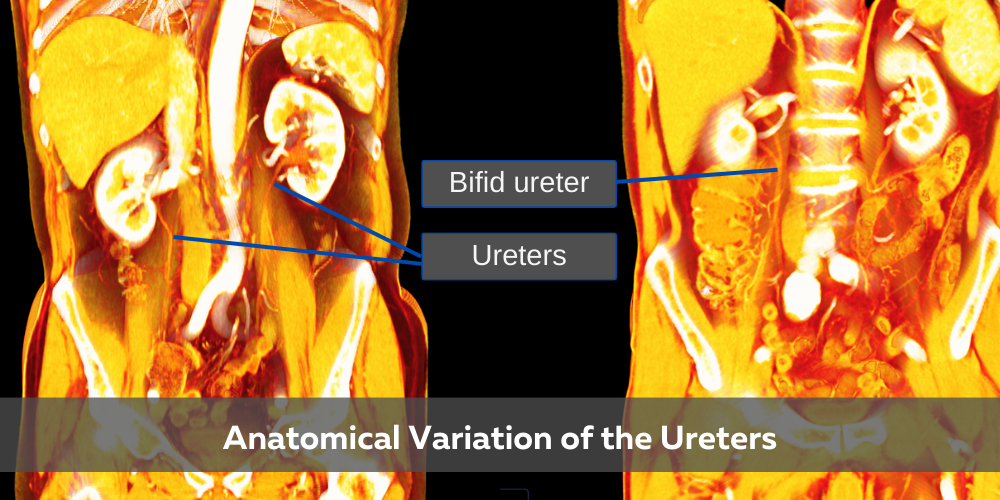
Surgical Consideration: Bifid Ureter
A bifid ureter, a split in the tube that carries urine from the kidney to the bladder, occurs in approximately 1 in 125 people. This variation can complicate urological surgeries and lead to recurring infections if not properly identified. BodyViz allows students to visualize this variation and understand its clinical implications.
Transplant Challenge: Accessory Renal Artery
An accessory renal artery, which is more common than you might think (occurring in up to 30% of people), can impact kidney transplants if not properly identified. By exploring these variations in 3D, students develop a more nuanced understanding of renal vascular anatomy.
These aren’t just academic examples, they’re real challenges that healthcare providers face every day. Helping students recognize and understand them early sets the stage for better clinical decision making.
Support and Implementation
Beyond the software itself, BodyViz is known for providing outstanding support to educators implementing the technology. This comprehensive approach ensures that institutions get the most value from their investment.
Dr. Bob Frampton shares his experience:
“The entire experience, from talking with Curt [CEO] to onboarding, was phenomenal. I’ve worked with a lot of other vendors, and the support from BodyViz is exceptional by comparison.”
Amy Wuthrich from DeSoto County High School also emphasized the ease of integration and the high level of support provided by the BodyViz team, noting that the software aligns well with state standards and made the transition smooth for incoming teachers.
Building the Future of Anatomy Education
As healthcare becomes more personalized and technology driven, anatomy education must evolve too. By teaching students to think critically about anatomy and not just memorize it, we’re helping them become better problem solvers, stronger clinicians, and more compassionate caregivers who understand that each patient’s anatomy is unique.
Amy Wuthrich summarizes this philosophy beautifully:
“It meets them where they are and gives them a real opportunity to succeed. BodyViz helps students understand human uniqueness at an anatomical level.”
See the Body Differently
Anatomical variations are more than medical curiosities; they're part of what makes us human, and understanding them is essential for effective, patient-centered care. With BodyViz, educators can bring real anatomy into the classroom and give students the tools they need to succeed.
The evidence is clear: BodyViz enhances understanding, improves retention, and better prepares students for clinical practice.
Are you ready to transform how your students learn about anatomical variations? Want to give them the advantage of exploring anatomical diversity through cutting-edge technology?
This article contains information adapted from "The Importance of Anatomical Variations" written by Dr. Robert Tallitsch. Originally published by BodyViz in November of 2022.