Creighton University School of Medicine
February 3, 2020
Innovation and technology is changing medical education in profound ways. The Creighton University School of Medicine recently embraced these changes after implementing a BodyViz solution to enhance the teaching of its anatomy classes.
“There have been a variety of technologies introduced in recent years that have attempted to bring the study of anatomy online,” said course director Dr. Ken Kramer, Assistant Professor in Biomedical Sciences and Medical Education. “Unfortunately, none of them were the complete solution we wanted.”
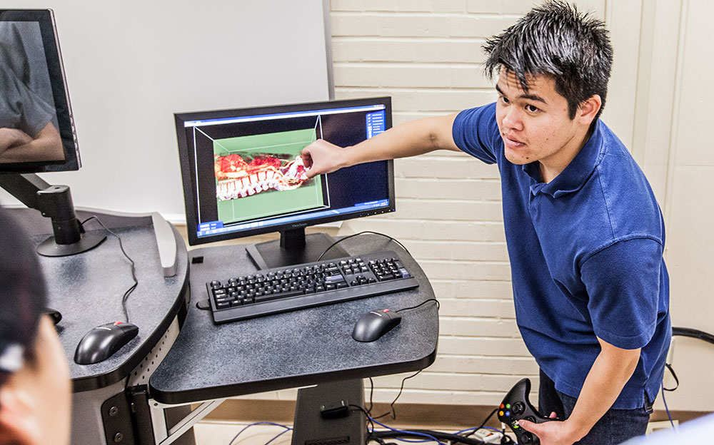
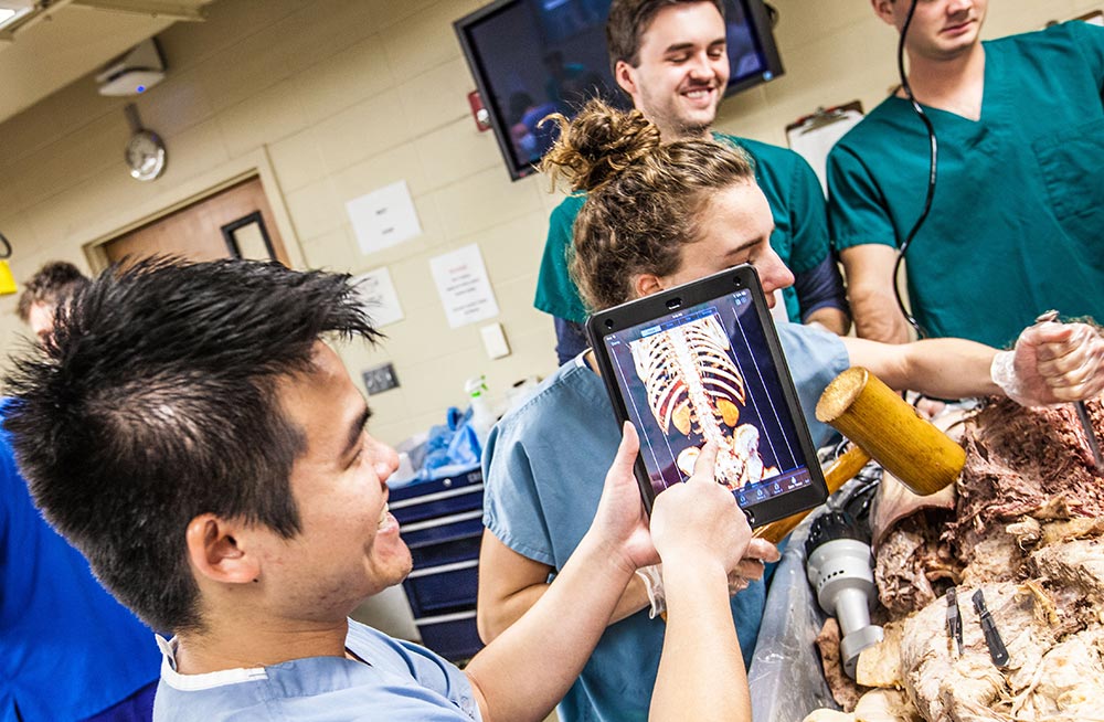
Dr. Kramer and his team were looking for a tool that enhanced the current curriculum, enabled students to view their cadaver scans during dissection, and allowed them to access a library of 2D and 3D images for comparison. He wanted all of this available in a 3D learning environment, as well as on iPads that would be used in the cadaver lab.
“There is variation in human anatomy even in what is considered normal. We can have 25 cadavers in a lab and there will be slight to significant variations in the basic anatomy. We can walk from table to table and discuss what is different, but it is difficult to compare what we’re seeing without the visuals in front of us. With the new technology, we can look at the cadavers in the room, then go next door to the BodyViz lab and compare the images from the students’ own cadavers to images we’ve selected from the library. Because the lab is always open, students can come in and revisit these scans on their own."
"Not only is this important for the current class, but it is key to preparing our students for what they will see in their clinicals and rotations."
The image library is a collection of thousands of real images that have been accumulated by BodyViz over the years through various donors and partnerships. Both students and educators can access the library from their iPads or the computer. The advantage of Creighton’s BodyViz lab is the ability to view real images - instead of models - in stereoscopic 3D.
“The images found in our library can be used and manipulated to suit a school’s current curriculum,” said Scott Rodenburg, BodyViz’s director of business development. “You can investigate using clipping planes, filter through various tissue densities, add labels, as well as drop the original and altered images into PowerPoint or a learning system. You can view them either as mono or stereoscopic images – depending on the BodyViz solution you have. All of this is giving the students and instructors unprecedented access to the human body and providing remarkable detail about spatial relationships in the body, unseen before.”
“In less than a semester, we’ve been able to update a tried-and-true method of teaching anatomy and bring it into the 21st century. It is providing our students with a different level of education that will provide a sound foundation for their future studies. We’ll definitely be looking at how we can expand this into other areas outside of our core anatomy curriculum.”
As Creighton University's School of Medicine begins using BodyViz, they have been pleased with the ease of implementation and the ability to incorporate the solution into their current lesson plans. Dr. Kramer noted they will look at scanning their own cadavers, and they plan on expanding the use of the technology.