A.T. Still University
November 4, 2019
Many of you can relate to the limitations and difficulties of running a cadaver lab. Although providing exceptional anatomical and dissection experiences, cadaver labs pose many restrictions for students and instructors regardless of academic level.
- Limited time and hands-on experience with anatomical resources
- Inability to consistently visualize variations, injury, disease, pathology, etc.
- Annual cost and regulations associated with obtaining and maintaining cadaveric specimens
Like many of you, the Anatomy Department at A.T. Still University, SOMA is all-too-familiar with these issues. The department chair, Dr. Jay Crutchfield, set out to modernize their lab resources to alleviate these issues for students and instructors, and teach clinically relevant anatomy and radiology without cadaver dissection as a requirement.
Dr. Crutchfield was intrigued by the many benefits of virtual anatomy software but was skeptical on how it would integrate with his courses that were in the middle of transitioning out of a dissection-based curriculum. The BodyViz team helped Dr. Crutchfield design a virtual anatomy solution that not only alleviated his curriculum from their cadaver lab frustrations, but also aligned directly with their individual curriculum goals.
"We needed to pick a solution that would help recruit students to our university and open up a modern venue for teaching radiology and cross-sectional anatomy, which is no doubt, the most necessary visual requirement for future physicians to become familiar with.
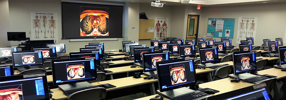
After fitting their anatomy classroom with 56 touch-screen computer stations, BodyViz provided ATSU students with significantly more access to hands-on anatomical resources. This helped Dr. Crutchfield create a curriculum where students are learning how to read medical imaging data of normal human anatomy, from head-to-toe, in a single year. With this advanced understanding, students are much more comfortable and prepared when transitioning into their clinical rotations.
Within ATSU's 3D Stereoscopic Virtual Anatomy Lab, students work individually at each of the 56 computer workstations where they virtually dissect and explore thousands of examples of real human anatomy. This hands-on approach familiarizes students with the complex 3D anatomical relationships and intricate details before stepping into the lab. At the front of the room, there is an additional workstation for the instructor. This workstation, however, is fitted with a 3D Stereoscopic Projector that displays lifelike 3-dimensional anatomical volumes when viewed using 3D glasses. These detailed visualizations are not only engaging, but they help students comprehend complex 3D spatial relationships more effectively.
Utilizing an existing learning environment, ATSU alleviated their anatomy curriculum from the limitations of a cadaver lab and increased their students' hands-on experiences with anatomical resources. To build upon the increased comprehension and confidence levels in his students, Dr. Crutchfield is hoping to further expand BodyViz access to all ATSU students using tablets.
To learn more about how BodyViz Virtual Anatomy Software can help you alleviate your students from traditional anatomy resource restrictions, schedule an interactive demo.