The Vertebral Column and Sciatica | 3D Anatomy Software
by Robert Tallitsch, PhD | October 8, 2021

Use this patient case study video created from a real patient example to help your students visualize the vertebral column and sciatica in 3D!
Written by: Robert Tallitsch, PhD
The vertebral column is part of the axial skeleton. It supports the head, protects the spinal cord, and serves as a point of attachment for the ribs and pelvis.
The vertebral column consists of individual bones, called vertebrae, and associated connective tissue composed of intervertebral discs and ligaments. There are 24 pre-sacral vertebrae: 7 cervical vertebrae, 12 thoracic, and 5 lumbar. There are two composite vertebrae in the pelvic area: the sacrum and the coccyx.
When referring to a particular vertebra the notation utilized by anatomists is a capital letter followed by a subscript number. The letter refers to the particular region of the vertebral column, while the number indicates the specific vertebra. For instance, T8 refers to the 8th thoracic vertebra.
Variations in the number of vertebrae in a particular region do occur. It is quite common to find these variations in individuals who have no history of back pain.
Cervical Vertebrae
The first, second, and seventh cervical vertebrae (C1, C2, C7) have unique characteristics, and are classified as atypical cervical vertebrae. The other cervical vertebrae are termed typical cervical vertebrae.
Typical Cervical Vertebrae (C3, C4, C5, C6)
The body of a cervical vertebra supports only the weight of the head and the vertebrae superior to it. Therefore, it is the smallest of all vertebrae. As you move caudally along the vertebral column the size of the vertebral bodies increase.
Typical cervical vertebrae possess a bifid (notched) spinous process. Laterally the transverse processes are fused to the costal processes. These two processes surround the transverse foramen, which encircles the vertebral artery and vertebral vein.
All vertebrae articulate with the vertebra superiorly and the vertebra inferiorly by the superior and inferior articular facets, which are found on the superior and inferior articular processes.
Atypical Cervical Vertebrae (C1, C2, C7)
The atlas (C1) has several unique characteristics:
- The atlas lacks a body.
- The atlas possesses the largest vertebral foramen of any vertebra.
- The vertebral foramen is surrounded by anterior and posterior vertebral arches.
The axis (C2) possesses a dens (odontoid process). During embryonic development the body of C1 migrates inferiorly and fuses with the body of C2, forming the dens. This structure, along with the transverse ligament that binds the dens to the inner surface of the atlas, allows you to turn your head from side to side.
C7, the vertebral prominens, has the longest spinous process of all of the cervical vertebrae. This vertebra marks the transition between the cervical and thoracic vertebral regions. It sometimes lacks a transverse foramen.
Thoracic Vertebrae
Each thoracic vertebra has a body that is larger than that of the cervical vertebrae, and a smaller vertebral foramen. The spinous process of each vertebra projects posteriorly and caudally. Each thoracic vertebra articulates with one or two ribs laterally. This articulation occurs at a costal facet, which is located on the anterolateral aspect of the body. The location and structure of these facets varies from thoracic vertebra to thoracic vertebra.
Lumbar Vertebrae
Lumbar vertebrae support the most weight of all of the vertebrae, and therefore are the largest vertebrae within the vertebral column. The spinous processes are short and stumpy, and project posteriorly. Costal facets and transverse foramina are lacking.
Sacrum
Shortly after puberty the five sacral vertebrae begin to fuse, forming the sacrum. Fusion is complete between the ages of 25 and 30. Prominent transverse lines demarcate the boundaries of the former separate vertebrae. The superior articular processes of the sacrum form synovial joints with the inferior articulating processes of L5. A sacral canal begins between the superior articulating processes of the sacrum and continues throughout the length of the sacrum.
The Coccyx
The coccyx is made up of 3 to 5 (typically 4) very small coccygeal vertebrae. These vertebrae begin to fuse around the age of 26. The first two coccygeal vertebrae possess transverse processes and have unfused vertebral arches. The coccyx is an attachment site for ligaments and a muscle that constricts the anal opening.
Intervertebral Discs
Intervertebral discs are composed of fibrous cartilage. In the adult, with the exception of C1 and C2, intervertebral discs are found between all cervical, thoracic, and lumbar vertebra, as well as between the 5th lumbar vertebra and the first sacral vertebra. These discs serve to maintain the proper spacing between the vertebrae, as well as assist in the transmission of force between adjacent vertebrae.
An intervertebral disc is composed of an outer ring of fibrous cartilage (the annulus fibrosus) and an inner gelatinous mass (the nucleus pulposus). A vertebral endplate covers the surface of the intervertebral disc.
Sciatica and the Vertebral Column
The term sciatica refers to pain that radiates from one’s lower back inferiorly through the buttocks, hips, thigh, and leg, typically on only one side of the body. This pain is due to compression of the sciatic nerve, often as a result of a bulging or displaced intervertebral disc within the lumbar region of the vertebral column.
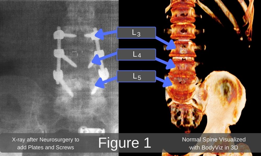
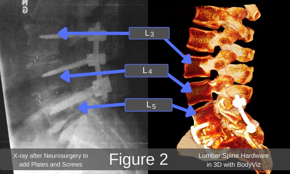
Figures 1 and 2 below are x-rays following neurosurgery on a patient in an attempt to increase range of motion and alleviate sciatic pain resulting from articular facet arthritis, spinal stenosis (a narrowing of the vertebral foramen) at L3, L4, and L5, and displacement of the intervertebral discs between L3 and L4, and between L4 and L5. As shown on the x-rays, screws and plates have been inserted to stabilize vertebrae L3 to L5.


Summary
The vertebral column consists of individual bones, called vertebrae, and associated connective tissue composed of intervertebral discs and ligaments. It supports the head, protects the spinal cord, and serves as a point of attachment for the ribs and pelvis. As with any part of the body, a pathology or malalignment often results in pain and an altered quality of life.
Want to learn more about 3D anatomy resources like the clips used in this BodyViz Brain Builder video and featured in this blog? Turn MRI and CT scans into real, 3D anatomy visualizations! The learning possibilities are limitless with our ever-growing library of patient case studies and anatomy scans. Excite and engage your students by bringing anatomy to life with BodyViz! Click the orange button to learn more about our 3D anatomy solutions that can fit your classroom's needs!
Learn More about 3D Anatomy Software
Helpful Links