The Benefits of Virtually Dissecting Real Cadavers with 3D Anatomy Software
October 1, 2020

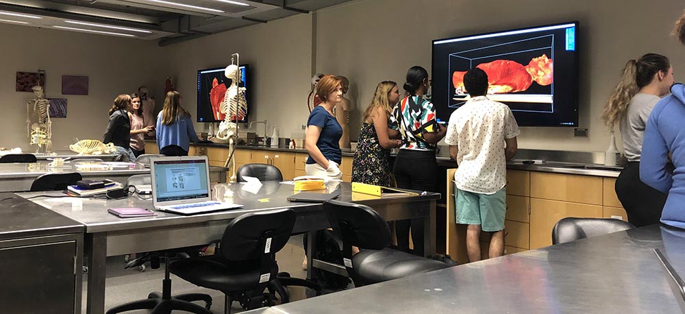
It’s no secret that 3D anatomy software has become increasingly popular within academic institutions as the need for more accessible learning resources became a requirement. Although a large portion of educators that have recently adopted these innovative platforms did so in response to online learning, there are a number of anatomy educators that have been utilizing 3D technologies in their virtual anatomy labs for years. Why? Because they are proven to be effective.
There is no doubt about it - cadaveric dissection is the best tool available when it comes to learning human anatomy and helping students empathize with death and dying; something that all medical professionals will encounter at some point in their careers. However, cadaveric dissection is just one of the many resources available for learning human anatomy and the 3D spatial relationships between structures. Students in the classroom today are far different from the students we had in our classrooms 15, 10, and even 5 years ago. These students, coming from the digital native generation, have proven to benefit most from anatomy courses while utilizing multiple pedagogical resources. In the article below, we will discuss a number of studies that reflect this claim. For additional resources, take a look at the related research and sources page on our website.
In a recent study, Mohamed Estai and Stuart Bunt review the range of teaching resources and strategies used in anatomy education with the aim of coming up with suggestions about the best teaching practices in this area. Their results show the best way to teach modern anatomy is by combining multiple pedagogical resources to complement one another, "students appear to profit most when diverse and system-based modalities are integrated." In a similar study performed by a team at the Oregon Health and Science University, led by Dr. Daren Nicholson, found that students with access to 3D anatomy software scored 18% higher in a post-module test compared to the control group that used traditional teaching methods. In line with Dr. Nicholson’s study, James Kulik’s research on computer-based instruction shows that students in health-related courses obtained a greater increase in test results when accompanied with computer-based learning, compared to the average among all other academic courses.
Dr Sonia Pujol from the Department of Radiology at Brigham and Women’s Hospital also supports this claim in her study titled, Using 3D Modeling Techniques to Enhance Teaching of Difficult Anatomical Concepts, where she explains that there is significant data showing that the interaction with 3D models led to a better understanding of the shape and spatial relationships among structures, and helped illustrate anatomical variations from one body to another. One student that participated in the study reported the following…
"The 3D imaging was really helpful in visualizing areas of the body that we could not see clearly in lab. Because the spatial relationships and surface anatomy are so important for clinical practice, the 3D imaging is such a useful learning tool and I hope that all of the areas of the body can be studied this way."
As anatomy curriculum goals grow denser and resources surrounding cadaver-based dissections decrease, a change in the way anatomy is being taught is inevitable. 3D anatomy software can flex to any teaching environment and scale to any pedagogical approach, and can do so in a very cost-effective manner. If you can provide your students with a more engaging resource, unlimited access to real anatomy in any location, and alleviate your institution from resource pressures, all while generating a better performance from your students - what’s holding you back?
Helpful Links