Carpal Tunnel Syndrome
by Robert Tallitsch, PhD | May 20, 2024
.jpg)
Video explaining Carpal Tunnel Syndrome with a patient example case study!
Use the button below to schedule a demo to learn about our activities, flash cards, and other anatomy resources that support this Brain Builder!
Schedule a Demo
The bones and muscles of the pectoral girdle and upper limb position the hand to accomplish a desired function. This desired function is then accomplished by the extrinsic and intrinsic muscles of the hand, which appropriately move the bones of the wrist, palm, and digits.
Carpal Tunnel Syndrome (CTS) is a common problem affecting hand function. CTS is caused by compression of the median nerve at the wrist. As a result of this compression, hand and wrist pain and other symptoms associated with carpal tunnel syndrome make movements of the hand and wrist quite problematic.
In this Brain Builder we will briefly discuss the muscles of the hand. Then we will discuss the anatomy of the carpal tunnel. Finally, we will discuss the symptoms, causes, and treatments for carpal tunnel syndrome.
Muscles of the Hand
The muscles of the hand are divided into two groups: extrinsic and intrinsic muscles. Extrinsic muscles of the hand have their origin on the bones of the forearm and insert into various locations of the wrist and fingers. These muscles provide strength and gross motor control of the hand and fingers. Two of these muscles, the flexor digitorum superficialis and the flexor pollicis longus, are innervated by the median nerve.
In contrast, intrinsic muscles of the hand originate on the carpal and metacarpal bones of the wrist and hand, and insert onto the phalanges of the hand. These muscles provide fine motor control of the hand and fingers.
Intrinsic muscles of the hand innervated by the median nerve are the:
-
Opponens pollicis
-
Abductor pollicis brevis
-
Flexor pollicis brevis
-
Lumbrical muscles
As the above lists show, the median nerve innervates several extrinsic and intrinsic muscles of the hand. These muscles allow for abduction and flexion at the wrist, flexion, abduction, and opposition of the thumb, flexion at the metacarpophalangeal joints, and extension at the proximal and distal interphalangeal joints.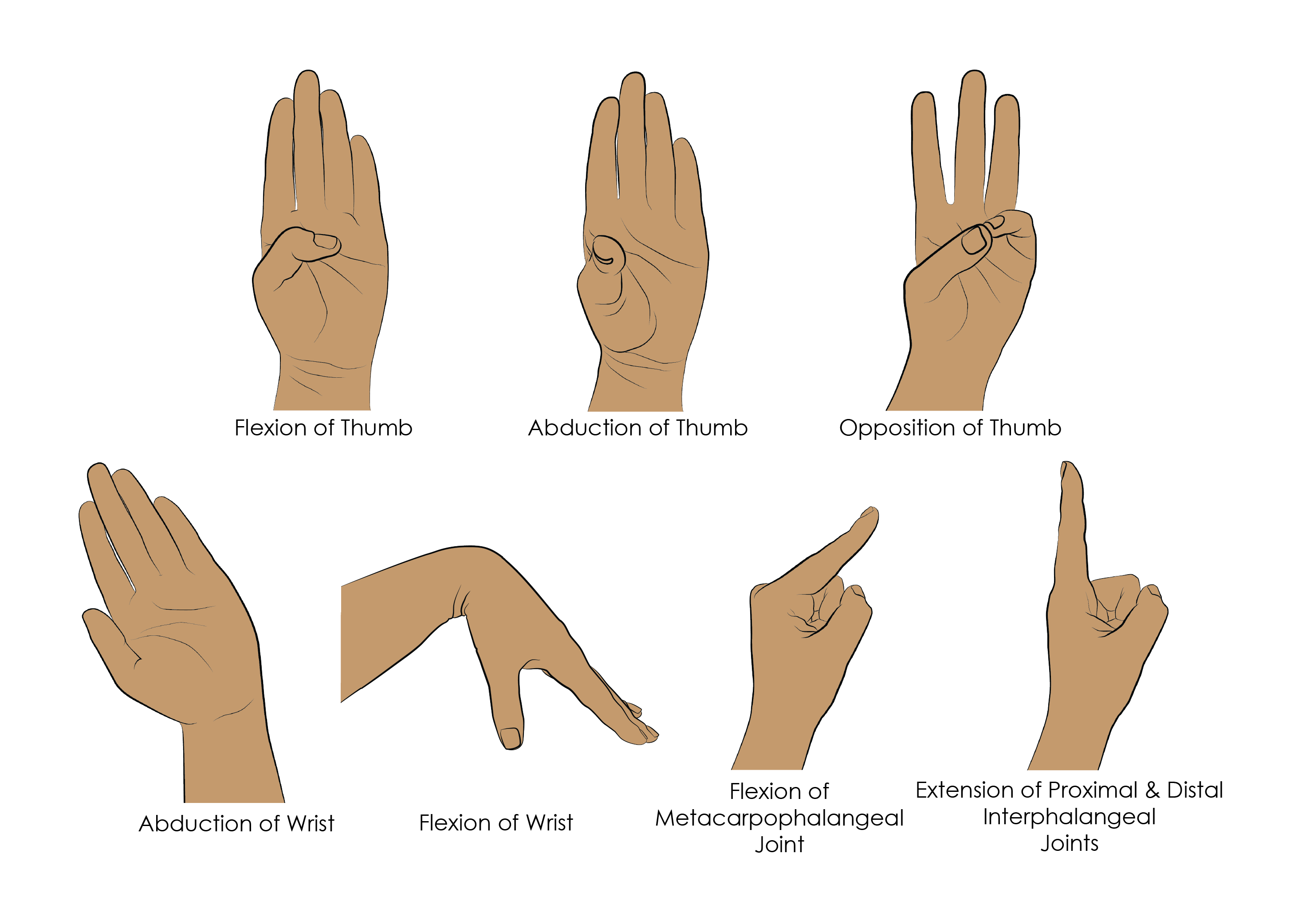
Anatomy of the carpal tunnel
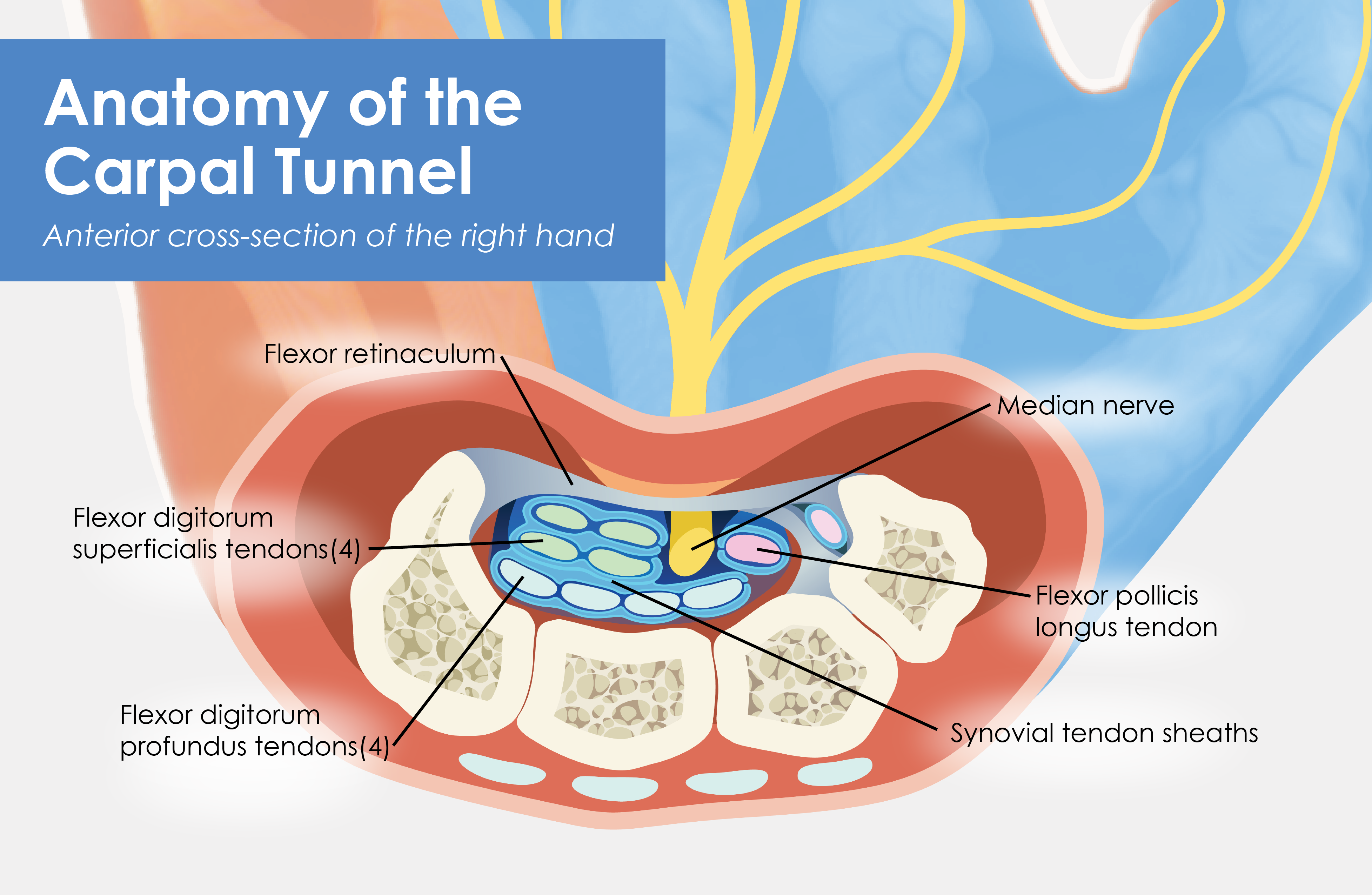
The carpal tunnel is two anatomical structures: (1) a deep arch of carpal bones on the anterior aspect of the wrist and (2) the flexor retinaculum. The carpal arch is formed by the pisiform and the hook of the hamate medially, and the tubercles of the scaphoid and trapezium laterally. The flexor retinaculum, which is a thick ligament, bridges the arch, connecting the medial and lateral sides of the arch, forming the carpal tunnel. The median nerve, the four tendons of the flexor digitorum profundus, the four tendons of the flexor digitorum superficialis, and the tendon of the flexor pollicis longus pass through the carpal tunnel.
Each of the nine tendons of the muscles mentioned above are enclosed within synovial tendon sheaths, which facilitate the free movement of the tendons within the carpal tunnel. All of the tendons of the flexor digitorum profundus and flexor digitorum superficialis are enclosed within a single sheath, while the tendon of the flexor pollicis longus is enclosed within its own, single sheath. The median nerve, which passes through the carpal tunnel anteriorly and slightly laterally to the muscle tendons, is not enclosed within a synovial sheath.
Symptoms of Carpal Tunnel Syndrome
As mentioned previously, the median nerve innervates extrinsic and intrinsic muscles of the hand, providing strength and both gross and fine motor control to the hand and fingers. In addition, the median nerve provides sensory input from digits one through three (thumb, index, and middle fingers) and the lateral half of the fourth digit (ring finger).
Symptoms of carpal tunnel syndrome are all associated with either the sensory or motor functions of the median nerve. Examples include, but aren’t limited to, the following:
-
Weakness of the thumb
-
Hand and wrist pain
-
Tingling and/or numbness of the hand
-
Reduced strength and motor control of the hand
-
Numbness, burning, and/or tingling in digits one through four
-
Perception of an “electric shock” through the wrist and hand
Often flexing the wrist enhances these symptoms, as this increases pressure on the median nerve within the carpal tunnel.
Causes of Carpal Tunnel Syndrome
One of the most common causes of carpal tunnel syndrome is the result of long-term repetitive actions, such as typing, texting, or playing the piano. These repetitive actions often cause swelling of the synovial membranes surrounding the four tendons of the flexor digitorum profundus, the four tendons of the flexor digitorum superficialis, and the tendon of the flexor pollicis longus. This places increased pressure on the median nerve, causing one or more symptoms listed above.
Other causes of carpal tunnel syndrome that may cause swelling of the synovial tendon sheaths include a fractured or dislocated carpal bone, obesity, pathology of the thyroid gland that alters the secretion of thyroid hormones, diabetes, and hormonal changes resulting from menopause or pregnancy.
Diagnosis and Treatment
Diagnosis of carpal tunnel syndrome will include one or more of the following procedures:
-
Detailed medical history that details when the pain starts and what motions or actions will increase or decrease the pain.
-
Assessment of hand strength, as well as strength and sensitivity of digits one through four.
If physical therapy and/or the use of a brace does not alleviate the pain the physician will utilize imaging and/or electromyogram and nerve conduction studies to determine whether or not surgery is recommended.
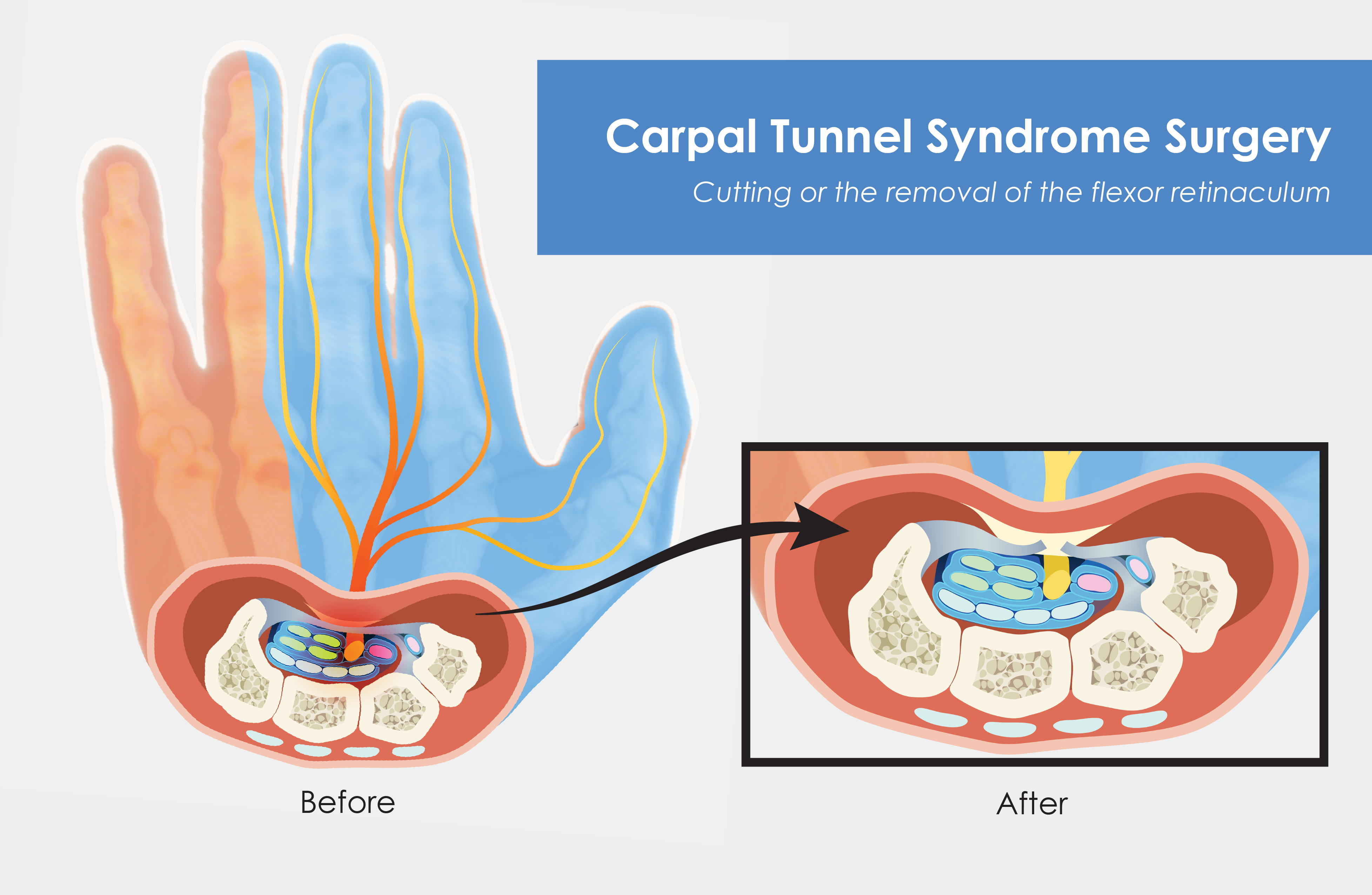
If surgery is recommended the physician will opt between open incision or endoscopic surgical procedures. Both of these procedures have the same goal: increasing the width of the carpal arch and carpal tunnel. This is accomplished by cutting some or all of the flexor retinaculum. The widening of the carpal arch and carpal tunnel reduces the pressure on the median nerve and, hopefully, lead to a resumption of normal functioning of the nerve.
Post-surgical treatment typically involves physical therapy, and the recurrence of symptoms of carpal tunnel syndrome is rare.
Key Terms
Flexor retinaculum - A thick ligament that bridges and connects the medial and lateral sides of the arch, forming the carpal tunnel.
Extrinsic muscles of the hand - Extrinsic muscles of the hand have their origin on the bones of the forearm, and insert onto various locations of the wrist and fingers. These muscles provide strength and gross motor control of the hand and fingers.
Carpal arch - An arch formed on the anterior surface of the wrist. The carpal arch is formed by the pisiform and the hook of the hamate medially, and the tubercles of the scaphoid and trapezium laterally.
Long-term repetitive actions - Actions such as typing, playing the piano, or texting that are done frequently and over a long period of time.
Intrinsic hand muscles - Intrinsic muscles of the hand originate on the carpal and metacarpal bones of the wrist and hand, and insert onto the phalanges of the hand. These muscles provide fine motor control of the hand and fingers.
Median nerve - One of the nerves of the forearm. The median nerve innervates four groups of intrinsic muscles of the hand, providing fine motor control to the hand and fingers. In addition, the median nerve provides sensory input from digits one through three (thumb, index, and middle fingers,) and half of the fourth digit (ring finger).
Function of the bones and muscles of the pectoral girdle and upper limb - The bones and muscles of the pectoral girdle and upper limb function to position the hand so that it can accomplish a desired function.
General function of the muscles whose tendons pass through the carpal tunnel - These muscles provide strength, gross and fine motor control to the hand and fingers.
Schedule a demo today to learn how you can incorporate BodyViz into your classes!
Schedule a Demo
Helpful Links